
X-ray
An X-ray is an electromagnetic radiation that has wa avelength shorter than ultraviolet rays but longer than gamma rays. The wavelength of the X-rays is approximately in the range of 10 nanometers up to 10 picometers, equivalent to the frequency range of 30 petahertz to 30 exahertz, or its photon energies range between 100 eV to 100 keV.

X-rays can penetrate many solid substances, such as construction materials and living tissue, and are widely utilized in radiography in medical diagnostics and material science, for example, to identify some chemical elements and detect weak points in construction materials. They are ionizing radiations. Their exposure is harmful to health as they damage DNA, cause cancer, dexa sacn, medical imaging, high intensities, burns, radiation sickness. Thus, their generation and usage are strictly controlled by public health authorities.
Observation and Test
X-rays were first discovered in science as a form of unknown radiation from discharge tubes by experimenters studying cathode rays generated by such tubes, which are high-energy electron beams first observed in 1869. Many of the early Crookes tubes exhibited effects that could be attributed to them. The vacuum tube produced free electrons by ionizing the residual air in the tube at a very high DC voltage ranging from a few kilovolts to 100 kV. This voltage accelerated the electrons released from the cathode at a high enough velocity that they developed X-rays upon striking the anode or the glass wall of the tube.
William Morgan is considered to be the first experimenter to produce successfully. He presented a paper to the Royal Society of London in 1785, about passing an electrical current through part of a glass tube that was evacuated and producing a glow created. Further work on this was carried out by Humphry Davy and Michael Faraday, his assistant at the time.
In 1888, Philipp Lenard performed experiments to determine whether cathode rays could pass from the Crookes tube into the air. He constructed a Crookes tube with a window at the end made of thin aluminum, facing the cathode so that the cathode rays hit it. He determined that something came through, which would expose photographic plates and cause fluorescence. He measured the penetrating power of these rays through different materials. It has been suggested that some of these Lenard rays were X-rays.
Continue in radiology
Taking an X-ray image with early Crookes tube gear, late 1800s. The Crookes tube is in the center. The standing man is looking at his hand on a fluoroscope screen. The seated man is taking a radiograph of his hand by placing it upon a photographic plate. No precautions against radiation exposure are taken since its hazards were not known at the time.
Röntgen soon realized X-rays had medical uses. With his 28 December Physical-Medical Society paper, he sent a letter to physicians he knew throughout Europe. News spread fast with Scottish electrical engineer Alan Archibald Campbell-Swinton being the first beside Röntgen to produce photograph. Over February, 46 experimenters were adopting the technology in North America alone.
The first clinical application was conducted by John Hall-Edwards in Birmingham, England on 11 January 1896, where he radiographed a needle lodged in the hand of an assistant. On 14 February 1896, Hall-Edwards was also the first to make use in a surgical operation.
Hazards discovered
With many stories of burns, hair loss, and worse following widespread experimentation with X-rays after their discovery in 1895 by scientists, physicians, and inventors came technical journals of the time. In February 1896, Professor John Daniel and William Lofland Dudley of Vanderbilt University tested hair loss after Dudley. In 1896, a child who was shot in the head came to Vanderbilt’s laboratory.
Before any attempt to locate the bullet, an experiment was attempted for which Dudley volunteered, true to his scientific nature. Daniel reported that, 21 days later, the photograph of Dudley’s skull, exposed for one hour, revealed a bald spot, 5 centimeters in diameter, on the part of his head nearest tube. A plate holder was fastened over his head with the plates toward the side of the skull, between the skull and his head was placed a coin. It was further fastened at the other end half-inch space from the hair. Apart from burns, alopecia, carcinoma, X-rays, radiation.
Soft and hard X-rays
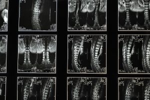
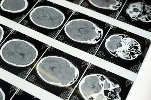
Hard X-rays are those that have a photon energy greater than 5–10 keV, while the lesser energy X-rays are referred to as soft X-rays. X-rays of a few keV in energy are sometimes called tender X-rays. The penetrating nature of hard makes them a common tool for radiographing objects and the contents inside them- for example, in medical radiography and airport security.
The term not only describes the method itself but is used metonymically to describe a radiographic image made with this method. Since the wavelengths of hard are comparable to the size of atoms, they also come in handy when ascertaining crystal structures through crystallography. Conversely, soft X-rays have attenuation lengths of less than a micrometer in water. The attenuation length of 600 eV in water is less than 1 micrometer.
Gamma rays
There is no consensus on the definition that splits from gamma rays. One convention makes a distinction based on the origin of the two types of radiation, originate from electrons, but gamma rays originate from the nucleus of the atom. This leads to several associated difficulties, other processes can produce these photons with very high energy as well, or sometimes it is not known how they originated.
Properties
The X-ray photons have sufficient energy to ionize atoms and break molecular bonds. Therefore, the radiation is ionizing and consequently destructive for the living tissue. Burning and radiation sickness in a very high dose over a short time frame and an increased risk of radiation-induced cancers for lower doses. Generally, the increased cancer risk from this examination is much outweighed by the benefits of the examination in medical imaging. Ionizing can be used for the killing of malignant cells by radiation therapy, apart from their application in the characterization of materials by spectroscopy.

CONCLUSION
Diagnostic X-rays increase developmental problems and cancer cases in people exposed to them. It is estimated that 0.4% of current cancers in the United States are due to computed tomography performed in the past and that this may increase to as high as 1.5–2% with 2007 rates of CT usage.
Experimental and epidemiological data presently do not support the proposition that there is a threshold dose of radiation below which there is no growing risk of cancer. However, this is under increasing doubt. At the dose of 1100 mGy, cancer risk may start coming into effect. The extra radiation from diagnostic is calculated to add an average person’s cumulative probability of developing cancer by age 75 by 0.6–3.0%. The absorbed dosage differs in various types of tests and body parts involved. Radiation dose for CT and fluoroscopy is relatively higher compared to plain X-rays.