
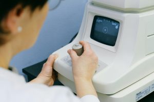
Optometry
Optometry is an allied health profession that entails the examination of the eyes and associated structures for defects or irregularities. Optometrists are health care providers who commonly offer full eye care.
Optometrists are professionals with a four-year Doctor of Optometry degree earned after they completed their undergraduate college education. Optometrists are licensed and educated to treat medical conditions related to the eyes aside from delivering refractive optical eye care. As part of their scope of practice, optometrists are deemed physicians and get reimbursed from medical insurance.
Optometrists can also give medical treatment prescribe drugs and conduct other operations for eye diseases besides giving refractive treatment. The Doctor of Optometry degree is less common in the UK. Optometrists engage a lot in scholarly research for eye related ailments and diseases. Besides prescribing spectacles and contact lenses for vision related deficiencies, optometrists are also trained in observing and managing ocular disease pathologies. The scope of training for optometrists also differs significantly between nations. There are some nations that only ask for certificate training, while others demand a doctoral degree.
Optometrists have a four-year college degree a four year Doctor of Optometry degree and are eligible to undergo a 1year residency program.
Optometry care
The treatment of age related macular degeneration was limited to restoring whatever vision had been lost due to the disease. Recent accomplishments of the anti vascular endothelial growth factor drugs and the antioxidant clinical studies have transformed the treatment of AMD. For the first time there is reasonable hope that the advancement of AMD can be halted or decelerated and near normal vision can be maintained.
Advances in new vision tests imaging modalities and genetic testing have significantly increased the likelihood of identifying the onset of AMD and choroidal neovascularization. But since existing treatments are still not able to restore degenerated retinal cells the most desirable patient outcome is early detection of the disease and implementation of proper treatment before substantial retinal damage has been inflicted. The potential and challenge posed by the novel treatment strategies and methods of detecting the disease have reshaped the role of primary care optometrists in managing AMD.
This literature review and practice guideline shows that beyond the classical functions of refraction and visual rehabilitation the distinctive role of optometrists as the primary eye-care professionals has enabled them to contribute significantly to the early detection of AMD patient education lifestyle change counseling disease monitoring and referral and nutrition supplement counseling.
The active involvement of primary care optometrists in shared care of AMD management will certainly lead to improved patient outcomes to a large extent. Optometrists must enhance their competence in these fields as well to respond to the new challenges. Though primary care optometrists have been dealing with patients with AMD all along their role in caring for this blinding disease has not been sufficiently documented.
Given the new game-changing advancements in AMD treatment it is timely to discuss what primary care optometrists are doing, what they can do, and what they ought to do to enhance patient outcomes in the era of new AMD management. Uveitis diagnosis is complex and there are diagnostic hints in both the clinical course and the clinical presentation.
Key of optometry
There are several key distinctions that need to be made to properly define the condition the Name Meshing System for diagnosing uveitis is helpful in providing a framework for categorizing these distinctions. With this structured framework uveitis treatment should include consideration of location duration pathology and laterality as well as presenting signs and symptoms.
Assessment of location is a critical first step in uveitis diagnosis. Anterior uveitis is a blanket term for iris anterior ciliary body or both inflammation. This represents a significant differentiation from intermediate or posterior segment uveitis regarding severity threat to vision and sequelae both of which are worse for the more posterior forms. Microscopically aqueous cells are diagnostic of iritis frequently with aqueous flare consisting of albumin. While anterior vitreous cells are evidence of cyclitis they are infrequently observed by themselves and are of limited clinical usefulness.
Another significant distinction in anterior uveitis is to recognize the acute course as opposed to the chronic course of the disease. Based on the Standardization of Uveitis Nomenclature criteria acute or limited cases imply an attack lasting <3 months whereas chronic or persistent cases persist beyond that period. This is a significant distinction clinically because the etiology and management approach differ between the two. The signs of chronic anterior uveitis are typically milder more frequently granulomatous in character and more likely to be associated with evidence of chronicity such as band keratopathy and iris changes like atrophy and lenticular changes. Recurrent anterior uveitis is defined as relapsing inflammation with >3 months of separation without treatment.
Infectious causes
Bacterial
Cat scratch disease is seen in immunocompetent persons of all ages globally. It is the most common cause of regional lymphadenopathy in children and young adults. The patient will have painful swollen lymph nodes at or near the location of the scratch or bite. It is the most frequent cause of neuroretinitis.
Lyme disease is a multisystem illness due to Borrelia bergdorferi spirochete infection and its complications transmitted by tick bites. In the USA the cases are localized in the northeast mid-Atlantic and upper midwest states. The rash is accompanied by fever, malaise, fatigue, arthralgia, and myalgias.
Several weeks after the initial exposure, the disseminated stage develops in the patients with skin, nervous system, joint, heart, and eye issues. Skin, nervous system and joints are involved in the chronic phase of this disease. Late neurologic manifestation appears as subacute or chronic encephalopathy with subtle cognitive and memory impairment progressive encephalomyelitis with white matter lesions and peripheral neuropathy. Uveitis can occur at the disseminated and chronic phases of the disease anterior uveitis intermediate uveitis posterior uveitis neuroretinitis, retinal vasculitis, choroiditis and panuveitis have all been described.

Viral
Viral infections are the most frequent infectious underlying cause of anterior uveitis. The IOP is elevated, iris atrophy occurs, and there are unilateral presentations with viral etiologies. Herpes simplex 1 and 2 are highly prevalent among humans in the world and contracted through direct exposure to active infection. In the majority of patients, this virus is latent in neural ganglia, and most notably the trigeminal ganglia.
Reactivation of the virus is triggered by stress, UV light, and illness or occurs in immunocompromised patients. Virus reactivation typically causes fever and malaise with skin vesicle eruption along the neural dermatome. Anterior uveitis caused by HSV is typically unilateral, granulomatous or nongranulomatous, and is associated with secondary elevation of IOP because of superimposed trabeculitis. It can also occur with intermediate uveitis, posterior uveitis, or panuveitis. Intermediate and posterior uveitis are treated with oral corticosteroids, antivirals, either orally or intravenously, and glaucoma drops.
Initial infection
The initial infection with varicella zoster virus human herpesvirus 3 is known as chicken pox, but following immunization with Varivax vaccine, cases of chickenpox in children became much lower in incidence after the mid-1990s. VZV lies dormant within neural ganglia and upon activation results in herpes zoster. HZ characteristically presents with unilateral pain along a dermatomal distribution in conjunction with a vesicular maculopapular rash. On reactivation along the trigeminal nerve it is called herpes zoster ophthalmicus.
It is also associated with an increased risk of autoimmune illnesses. Uveitis is commonly encountered during initial infection in IM. Human herpesvirus 5 cytomegalovirus is present in humans and may confer significant morbidity and mortality on immunocompromised hosts. CMV retinitis is the most prevalent presentation, presently rarely encountered in HIV/AIDS patients with the current common usage of highly active antiretroviral therapy. In a few cases immunocompetent patients can have unilateral, hypertensive, chronic, or recurrent uveitis secondary to CMV.