
MRI
Magnetic resonance imaging MRI is a form of radiological imaging through which images of the body can be taken. In the case of MRI scans images are created through strong magnetic fields and magnetic field gradients with radio waves delineating the organs of the human body. It differs since it doesn’t involve any ionizing radiation, unlike what happens in CT and positron emission tomography. It uses nuclear magnetic resonance, a sensitive instrument whose applications are not limited to imaging in various NMR applications. It includes among others NMR spectroscopy.
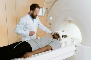
MRI is highly applied in hospitals and clinics to help diagnose, document and monitor disease cases. MRI images demonstrate better contrast between tissues than those obtained by CT scan, for example between the skin and the white matter of the brain or abdominal organs. But patients will find this less comfortable, as the process, which usually takes much longer and is also much noisier, requires the subject to lie in a long, narrow tube, although open MRI designs generally alleviate this problem.
Other metals, which do not move in the body, such as implants, are potentially harmful and can make it impossible for a few patients to have an MRI safely.
The term originally known as Nuclear Magnetic Resonance MRI was renamed to drop the word nuclear because of its connotations. Some atomic nuclei absorb RF energy if those nuclei are placed in an external magnetic field spin polarization induced in this way can induce an RF signal in a radio frequency coil and thus be detected. That is, a nuclear magnetic spin of protons in hydrogen nuclei resonates with radio waves and coherent radiation traveling in a compact direction, frequency, and phase of their energy.
Diagnostic MRI
Using organs or systems
The use of MRI has a variety of applications in medical diagnostics, and it is estimated that there are at least 50,000 scanners in use today. Even though the impact of MRI applications in diagnosis and treatment in many specialties can clearly be seen, the effect on improved health outcomes remains somewhat controversial in other instances.
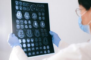
Images of the head and neck
MRI is the test of choice in the preoperative staging of rectal and prostate cancer and plays a role in the diagnosis staging and monitoring of others. Tumors also define areas of tissue for biobank sampling.
Neuroimaging
MRI is the investigative tool of choice for neurological cancers over CT because it allows better visualization of the posterior cranial fossa containing the brainstem and cerebellum. It has emerged that MRI has become the best solution for most central nervous system conditions, such as demyelinating diseases, dementia, cerebrovascular disease, infections, Alzheimer’s disease, and epilepsy, chiefly because of gray and white matter contrast.
Given the fact that Multiple images are taken in milliseconds, showing how the brain responds to different stimuli, allows researchers to study functional and structural abnormalities of the brain in psychological disorders. MRI is also used in surgery and stereo-toxically guided radiosurgery in the treatment of intracranial tumors, arteriovenous malformations, and other conditions that can be treated surgically with a device known as N-localizer. New tools integrating artificial intelligence in healthcare have enabled better image quality and morphometric analysis in neuroimaging through the use of a denoising system.
Cardiovascular
MRI of the heart is an adjunct to other imaging modalities. It is particularly useful for the anatomical and functional rating of the heart. Applications include myocardial ischemia and stability, cardiomyopathies, myocarditis, iron overload, vascular disease, and congenital diseases.
Angiography
MRA generates pictures of the arteries to look for stenosis, narrowing, or other abnormalities, as well as aneurysms in the walls of blood vessels, and even ruptures. MRI is most commonly used to examine the arteries in the neck and brain, also including the thoracic and abdominal aorta, renal arteries, and legs. Several methods can be used to produce the images, such as the administration of a paramagnetic contrast agent, like gadolinium, or flow contrast, in which most of the signal in an image is provided by blood flow in that plane.
Techniques involving phase binning, termed phase contrast angiography, may also be readily applied to produce accurate flow velocity maps.
Effects of MRI
The time taken by MRI is more than the time taken by CT. Furthermore, MRI is relatively less accessible than CT. This is the reason why CT scanning might be a more favorable option in such emergent or severe conditions like stroke. MRI scanning is also costlier than CT scanning.
Space Issues
The inside space of the MRI scanner is confined and rather small in size. Some people feel claustrophobic even though they don’t mind enclosed spaces. Sometimes, obese individuals cannot fit into a scanner. There are open MRI scanners. They are called such not because they are open in the front and back but because one side of their interior is an opening. Obese people are not bothered and get accustomed to it easily.
The images taken in open scanners depend on the strength of the magnet and are generally of lower quality compared to those produced by closed scanners but the diagnosis will not be difficult to establish. Anxious patients can be treated with an anxiolytic medication such as alprazolam 15 to 30 minutes before the exam.

MRI Safety
MRI in general is a safe procedure, except for injuries because of neglected safety procedures or human error. Most cochlear implants and pacemakers are contraindications to MRI, as well as metal fragments and foreign bodies within the eye. Magnetic resonance imaging during pregnancy appears to be safe at least during the second and third trimesters if performed without contrast agents. MRI does not use ionizing radiation. The amplitude and rate of change of the gradient coils during image acquisition may cause peripheral nerve stimulation.
External video
Video icon Magnetic objects thrown into an MRI magnet MRI uses powerful magnets and therefore magnetic materials move at very high speeds, leading to a projectile hazard and possibly fatal accidents. Deaths are very rare, however, with over a million MRIs performed each year worldwide.
MRI machines generate noise of 120 decibels. This can lead to hearing loss. So all individuals in the MRI room have to wear appropriate hearing protection at the time of examination.