
Angiography
Angiography or arteriography is the medical imaging of the lumen of the body’s blood vessels and organs. It is taken mainly through the arteries, veins, and heart chambers. Modern angioplasty is done through the injection of a radio-opaque contrast agent into the blood vessel and imaging by using X-ray-based techniques, which include fluoroscopy. The movie or picture of the blood vessels is called an angiograph, or more commonly an angiogram. Although it may be used for both arteriogram and venogram, the terms used most often are angiogram and arteriogram for the former, and venogram is only used for the latter.
Types of Angiography
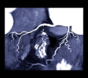
Cerebral angiography
Cerebral angiography is an angioplasty that aids in producing images of blood vessels in and around the brain, thereby detecting abnormalities such as arteriovenous malformations and aneurysms. The first to pioneer it was the Portuguese neurologist Egas Moniz in 1927 at the University of Lisbon. He was also involved in developing thorotrast for this procedure.
Usually, the catheter is inserted in a large artery, such as the femoral artery, and then passed through the circulatory system to the carotid artery, where the contrast agent is introduced. Several radiographs are then taken while the contrast agent distributes itself within the brain’s arterial system and a second set of radiographs is when the contrast agent reaches the venous system.
Applications of Cerebral Angiography
Cerebral angiography is used for diagnosis but is sometimes followed by treatment procedures in the same setting. It is used to image a variety of intracranial within-the-head or extracranial outside-the-head diseases.
In addition, the indications include non-traumatic subarachnoid hemorrhage, non-traumatic intracerebral hemorrhage, intracranial aneurysm, stroke, cerebral vasospasm, cerebral arteriovenous malformation for Spetzler-Martin grading and plan for intervention, dural arteriovenous fistula, embolization of brain tumors like meningioma, cavernous sinus haemangioma, for Wada test, and to obtain hemodynamics of cerebral blood flow such as cross-flow, circulation time, and collateral flow.
This also includes subclavian steal syndrome, ruptured carotid artery, carotid artery stenosis, cervical spine trauma, epistaxis nose bleeding, and an embolization plan of juvenile nasopharyngeal angiofibroma preop.
Pulmonary angiography
Pulmonary angiography is a medical fluoroscopic procedure that is used in the visualization of the pulmonary arteries and much less commonly, the pulmonary veins. It is a minimally invasive procedure most often performed by an interventional radiologist or interventional cardiologist to visualize the arteries of the lungs.
Uses of pulmonary angiography
Pulmonary angiography is useful as a diagnostic tool when non-invasive imaging such as CT pulmonary angiography is negative to rule out the diagnosis of pulmonary embolism. Pulmonary angioplasty might be more sensitive than clinical assessment, arterial blood gas, and ventilation-perfusion scan. Pulmonary angioplasty can also be used in the diagnosis of chronic thromboembolic pulmonary hypertension CTEPH and it can serve as a basis for balloon pulmonary angioplasty to treat the disease.
CT pulmonary angioplasty has become the method of choice for conventional pulmonary angioplasty in daily practice nearly exclusively because it is less invasive, quicker, safer, and delivers most of the same information with the bonus of demonstrating lung tissue and other structures. It is used, however, when CT angioplasty is not diagnostic.
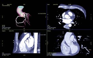
Radio angiography
This is a sub-specialty in nuclear medicine concerning imaging aimed at assessing the functionality of the right and left ventricles of the heart. This thus facilitates an educated intervention in the case of heart failure. It uses a radiopharmaceutical for injection into the patient, a gamma camera for acquiring, and a MUGA scan involving the acquisition of a cardiac cycle triggered at multiple points, among others. MUGA scanning is also known as equilibrium radionuclide angiocardiography, radionuclide ventriculography RNVG, gated blood pool imaging, and SYMA scanning, which is synchronized multi-gated acquisition scanning.
Purpose of Radionuclide ventriculography
Evaluation of coronary artery disease, valvular heart disease, congenital heart diseases, cardiomyopathy, and other cardiac disorders Radionuclide ventriculography is also performed. MUGA is usually prescribed for patients after the following conditions Known or suspected coronary artery disease diagnosis of the disease and prognosis of its outcomes Lesions in their heart valves congestive heart failure
People who have undergone percutaneous transluminal coronary angioplasty, coronary artery bypass graft surgery, or medical therapy, to measure the effectiveness of the treatment
Those with low cardiac output following open-heart surgery, people undergoing cardiotoxic drug agents such as chemotherapy with doxorubicin or immunotherapy, and people with a cardiac transplant.
Benefits of Angiography
Heart and vascular CT angiography may offer surgery as an alternative. If, after imaging, surgery becomes necessary, it will then be even more precise.
Heart and vascular CT angioplasty may permit the stenosis or occlusion of blood vessels that can become a condition for potentially corrective treatment to be performed.
CT angioplasty could produce more detailed anatomy compared to other imaging, at least regarding smaller blood vessels.
Heart and vascular CT angioplasty is accessible to almost all patients diagnosed with problems concerning blood vessels. Vascular and heart CT angiography is faster, non-imposing, and has fewer complications.
It is a good means of identifying arterial for example, narrowing of blood vessels in the heart and venous disease, as well as structural abnormalities of the heart before there are symptoms, or when symptoms are not related to blood vessel disease.
There is potentially less discomfort because contrast material is injected into an arm vein.
No radiation remains in a patient’s body after vascular CT angioplasty.
Routine X-rays used for routine CT scans have no immediate side effects.
Problems
Angiography is a relatively safe process. However, it has a few minor and very rare complications. After an angiogram, acute shock can cause slight pain over the surgery area, although heart attacks and strokes usually occur not, as they probably would in bypass surgery. This risk of complications from an angioplasty can be minimized by earlier knowledge of the number of clots and their positional placement by using a scan by CT.
Other dangers
The contrast dye used also leaves a warm feeling usually that lasts only for a few seconds but might be even stronger in the region into which it was injected. Much worse complications are unavoidable if the patient is allergic to the contrast medium, however, new contrast agents make it very improbable a dangerous reaction to occur over one in 80,000 studies.
Along with that, vascular injury could be done at the level of puncture injection, as well as anywhere in the vessel along the pathway by the catheter. If digital subtraction angiography is used, then the risks are minimized as the catheter need not be advanced to that level in the blood vessels, thus minimizing the possibility of damage or blockage.
Infections
Antibiotic prophylaxis can be given in procedures that are not clean or clean procedures that produce a generation of infarcted or necrotic tissues such as embolization. Routine diagnostic angiography is usually taken to be a clean procedure. Prophylaxis also helps in preventing infection from an infected space into the bloodstream.