
Echo-cardiogram
An echocardiogram echo is a graphical tracing of your heart motion. Your doctor uses ultrasound high frequency sound waves from a hand held wand placed on your chest to photograph your heart valves and chambers during an echo test. This assists the doctor in assessing the pumping function of your heart. Providers usually pair echo with Doppler ultrasound and color Doppler methods to assess blood flow through your heart valves. Echocardiography involves no radiation. This distinguishes an echo from other tests such as X rays and CT scans that involve small doses of radiation.
It is commonly applied in the diagnosis, management and treatment of patients with any presumed or diagnosed heart diseases. It is among the most common diagnostic imaging modalities used in cardiology. It can yield a tremendous amount of useful information such as the hearts internal chamber size quantification, pumping ability, location and extent of any tissue injury, and valve assessment. An echocardiogram also provides doctors other measurements of cardiac function, including an estimation of the cardiac output, ejection fraction, diastolic function and how the heart relaxes.
They plays a crucial role in the evaluation of wall motion abnormality in patients with suspected cardiac disease. It is a diagnostic tool used to arrive at an early diagnosis of myocardial infarction, with evidence of regional wall motion abnormality. It also plays a significant role in the treatment and follow-up of patients with heart failure by measuring ejection fraction.
Uses of Echo
Health societies suggest the employment of echocardiography in initial diagnosis when there is a change in the patient’s clinical status and when new information from an echocardiogram would cause the physician to alter the patient’s treatment. Diagnostic criteria for many cardiac diseases are derived from echocardiography studies. The discrimination of mild, moderate and severe valvular disease is derived from measured criteria. Another is the estimation of cardiac function by the left ventricular ejection fraction LVEF with enormous applications such as heart failure classification and cut-offs for implantable cardioverter-defibrillator implantation.
Routine testing is not recommended by health societies when the patient does not have a change in clinical status or when a physician will not alter care for the patient based on test results. An example of routine overuse of echocardiography when not warranted is the employment of routine testing based on a patient’s diagnosis of mild valvular heart disease.
Patients in such instances are usually asymptomatic for many years before the deterioration sets in and the echocardiogram result would not lead to a change in management without an accompanying change in clinical status. Echocardiography plays a wide role in pediatrics, diagnosing patients with valvular heart disease and other congenital defects. A new branch is fetal echocardiography, which is the echocardiography of an unborn fetus.
Types of Echo
There are three main types of echocardiography
transthoracic, transesophageal, and intracardic. Stress testing uses transthoracic echo with an exercise modality.
Transthoracic echo
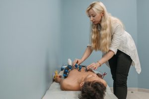
A routine echocardiogram is also referred to as a transthoracic echocardiogram or cardiac ultrasound, and it is employed for quick assessment of a patient at the bedside. Here, the echocardiography transducer is positioned on the subject’s chest wall and images are obtained through the chest wall. This is a non-invasive, highly sensitive, and rapid evaluation of the overall function of the heart.
TTE employs several windows to visualize the heart from various angles. Every window is better or worse for visualizing certain structures within the heart and usually, many windows are used in one study to evaluate the heart completely. Parasternal long and parasternal short axis windows are obtained alongside the sternum, the apical two three four chamber windows are obtained from the apex of the heart and the subcostal window is obtained from below the edge of the last rib.

Transesophageal echo
A transesophageal echocardiogram is a different method to conduct an echocardiogram. A dedicated probe having an ultrasound transducer at its distal tip is inserted through the mouth into the patient’s esophagus, enabling image and Doppler assessment from directly behind the heart. It is used most commonly when transthoracic views are poor and a clearer, more accurate view is required for evaluation. This procedure is conducted in the company of a cardiologist, anesthesiologist, registered nurse, and ultrasound technologist. Conscious sedation or local numbing medication can be employed to make the patient more comfortable throughout the procedure.
TEE, in contrast to TTE, lacks distinct windows through which to see the heart. The entire stomach and esophagus are available, and the probe being moved or withdrawn along this axis to change the view of the heart. The majority of probes have the facility to deflect the probe tip in one or two dimensions to further optimize the view of the heart. The ultrasound crystal is also usually a two-dimensional crystal and the plane of ultrasound being utilized can be electronically rotated to allow for an extra dimension to allow optimal views of the heart structures. Movement in all of these dimensions is usually required.
Stress echo
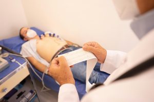
A stress echocardiogram or stress echo is a test that employs ultrasound imaging of the heart to compare the wall motion with physical stress. Images of the heart are initially captured when the patient is at rest to obtain a baseline of the patient’s resting heart rate wall motion. The patient subsequently walks on a treadmill or another modality of exercise to elevate the heart rate to his or her target heart rate or 85% of the predicted maximum heart rate of 220 minus the patient’s age.
Last but not least, images of the heart are acquired at stress to evaluate wall motion at maximal heart rate. A stress echo evaluates the heart wall motion, but it will not directly generate an image of the coronary arteries. Ischemia of the single or multi-coronary arteries may be responsible for the wall motion abnormality, potentially representing coronary artery disease. Cardiac catheterization is the gold standard for directly generating a picture of the coronary arteries and directly evaluating stenosis or occlusion. A stress echo is non-invasive and is conducted in the presence of a qualified medical practitioner a cardiologist and a cardiac sonographer.