
CT scan
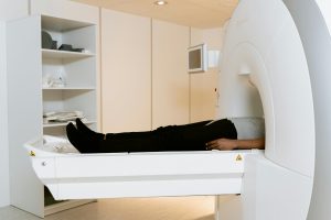
The procedure is clinically known as a CT scan which formerly was a CAT scan to give detailed impressions of the interior structures within the human body. Generally, those operators who offer the service for CT scans are called either radiographers or radiologic technologists
A rotating X-ray tube and a row of detectors placed inside a gantry scan X-ray attenuations by various tissues within the body. Multiple x-ray measurements taken from different angles are then processed on a computer with tomographic reconstruction algorithms to produce a series of tomographic images of a body. CT scans can be performed in patients possessing metallic implants or pacemakers, for whom MRI is contraindicated.
Since its invention in the 1970s, Computer tomography scanning has been an extremely versatile imaging technique.
Allan MacLeod Cormack and British electrical engineer Godfrey Hounsfield for the invention of computer-assisted tomography.
Types of CT scan
Sequential CT scan
It is known sequentially as step-and-shoot CT. In this scanning mode, the table steps that is, the table increments to a certain place and then holds, where the X-ray tube rotates during the acquisition of a slice. The table again increments while acquiring the next slice. Slices are acquired without stopping table movement. This takes more time to scan.
Spiral CT
CT scan of the thorax. Spinning tube, commonly called spiral CT, or helical CT, is an imaging technique in which an entire X-ray tube is spun around the central axis of the area being scanned. These are the most prevalent type of scanners in use because they have been around longer and cost less to produce and buy. The primary limitation of this type of CT is that the weight and inertia of the equipment X-ray tube assembly and detector array on the opposite side of the circle which limits the speed at which the equipment can spin. Some designs use two X-ray sources with detector arrays offset at some angle to improve temporal resolution.
Electron beam tomography
Electron beam tomography is a version of Computer tomography where the X-ray tube is built to be large enough that only the electrons, traveling from the cathode to the anode within the X-ray tube are spun using deflection coils. This type has a significant advantage because sweep speeds can be much faster, which will allow for less blurry imaging of moving structures, such as the heart and arteries. Fewer scanners of this design have been produced compared to spinning tube types, mainly due to the higher cost associated with building a much larger X-ray tube and detector array and limited anatomical coverage.
Dual Energy CT
Such a type of computed tomography is called Dual Energy CT or Spectral CT. In this advancement of computed tomography, two energies are deployed to prepare two sets of data. A dual-energy CT may use a dual source, a single source with a dual detector layer, and a single source with energy-switching methods to acquire two different sets of data.
Dual source CT represents the next generation of scanners, which employs a two X-ray tube detector system, rather than the conventional single tube systems. The two detector systems are mounted at 90° on one gantry in the same plane. Dual Source Computer tomography scanners provide high temporal resolution due to fast scanning as one full CT slice can be acquired within half a rotation. Fast imaging results in less motion blurring especially at high heart rates and potentially for shorter breath-hold time.
It is of particular benefit when the patients are ill, cannot hold their breath, or cannot take heart-rate-lowering medication.
Single Source with Energy switching is another version of Dual-energy CT wherein a tube is operated at two different energies by making repeated changes in the energies.
CT perfusion imaging
CT perfusion imaging is a modality of CT that is used to study flow through vessels as a contrast agent is injected. Blood flow, blood transit time, and organ blood volume can all be estimated with reasonable sensitivity and specificity. Such CT scan may therefore be applied to the heart, even though sensitivity and specificity remain lower compared to other modalities of CT. This can also be applied to the brain, where CT perfusion imaging can sometimes identify poor brain perfusion long before it has been identified via a conventional spiral CT scan. This is better for stroke diagnosis than other types of CT.
Medical application of CT scan
CT has become an important medical imaging tool in supplementing conventional X-ray imaging and medical ultrasonography. It has only recently been applied to preventive medicine or screening for disease, such as a CT colonography for high-risk individuals with colon cancer, or full-motion heart scans for high-risk, people with heart disease. A few institutions make full-body scans available to the public at large though this is an anathema to the recommendation and official position of most professional bodies in the field partly due to the dose of radiation involved.
CT scans have increasingly been used over the past two decades around the world. About 72 million were done in the United States in 2007, and more than 80 million in 2015.

Industrial use of CT scan
Industrial CT scanning industrial computed tomography is the process that uses X-ray equipment to produce 3D representations of components both internally and externally. Industrial CT scanning has been used in many industries for internal inspection of components. Some of the key uses for CT scanning have been flaw detection, failure analysis, metrology, assembly analysis, image-based finite element procedure, and reverse engineering applications. CT scanning is also working in the imaging and conservation of museum artifacts.
Aviation security use
CT scanning has also been applied in transport security largely within airport security, where it is already being used in a materials analysis context for explosives detection CTX and is also being considered for automated baggage parcel security scanning using computer vision-based object recognition algorithms that focus on the detection of specific threat items based on 3D appearance. Its use in the security of airports was pioneered at Shannon Airport in March 2022 a move that ended the ban on liquids over 100 ml there, a move that Heathrow Airport plans for a full roll-out on 1 December 2022, and the TSA spent $781.2 million on an order for over 1,000 scanners, ready to go live in the summer.
Geological use of CT scan
In geologic studies, X-ray CT is used to depict what is inside the drill core in a very short time. The dense minerals such as pyrite and barite appear bright and less dense clayey constituents appear dull, in CT images.
Paleontology use
Traditional methods of studying fossils are mostly destructive, such as thin sections and physical preparation. Non-destructive visualization of fossils in three-dimensional space is obtained by applying an X-ray CT scan to paleontology. This offers many advantages. For example, we can view fragile structures that may never be able to be studied otherwise.
utilization of cultural heritage
X-ray computer tomography and micro-CT can also be used to conserve and preserve objects of cultural heritage. Many fragile objects require direct research and observation that is damaging and also deteriorate the object in the long run. Using CT scans, conservators and researchers can determine the material composition of the objects they are examining good example is ink position along the layers of the scroll-without causing further damage. Even though they are not appropriate for every artifact to which such research questions could be applied, there are some artifacts, the Herculaneum papyri for example, in which the material composition varies very little along the inside of the object.

Advantages of CT scan
CT scanning has several advantages over conventional two-dimensional medical radiography. First, CT removes the superposition of images of structures outside the area of interest. Second, CT scans have a higher image resolution, allowing for the viewing of finer details. CT can differentiate between tissues with a difference of just 1% or less in radiographic density. Third, CT scanning allows for multiplanar reformatted imaging scan data can be viewed in the transverse, coronal, or sagittal plane according to the task of diagnosis.
CT has allowed for the development of new studies. For example, the angiography is performed by removing the necessity of placing an invasive catheter. CT scanning can now perform a virtual colonoscopy better and with less discomfort to the patient than an actual colonoscopy. Virtual colonography is far more sensitive than a barium enema in order to detect tumors and will require a reduced dosage of radiation.
CT is an intermediate to high-level radiation diagnostic test. Factors to determine the required radiation dose for a particular exam include volume scanned, patient build, number and type of scan protocol, and desired resolution and image quality. Two helical CT scanning parameters are easy to adjust and significantly affect the quantity of radiation tube current and pitch.